Hey guys, sorry for the late post...anyways, here's what we did in class yesterday (as today was a BIO DAY!!! I mean c'mon how interesting was that? "I had a little birdy, his name was Enza, I opened up the door and influenza"...haha so weird!!!!))
First we started off by turning in our multi-flow map and since Mrs. Stein's computer was down, we skipped the star&wish for Michael B's blog. Make sure to check out his blog and to comment on it.
Then when began to take notes in class, and I titled mine "Cancer Notes"
Here's how they looked:
Plant cells:
-Cell plate forms between cells.
-No centrioles.
-Spindle fibers
-Stages of Mitosis (PMAT)*
Animal cells:
-Centrioles
-Cell pinches at cleavage furrow.
(SAME AS PLANT CELLS):
-Spindle fibers
-Stages of Mitosis (PMAT)*
*Note: PMAT stands for the four stages of Mitosis; prophase, metaphase, anaphase, and telophase.
Then we went on to try to understand PH, which can be found in your text book on page 43-44.
Notes involving PH:
Acid is at a PH level at 14, a neutral is at a PH level 7, and a base is at a PH level of 14.
Examples: acid: stomach acid, soda; neutral: pure water; base: soap.
-If more positive charges than negatives, then it's classified as an acid.
-If it has more negative charges, then it's classified as a base.
We moved onto another section of notes: regulating the cell cycle, which is what our previous HW assignment was on; here are some notes:
-Cyclins are proteins that regulate the cell cycle.
-Spindle formation is during Interphase.
Ask yourself this question: What does DNA control?
And answer with this (one): DNA contains the instructions for making important proteins.
Note: If this goes wrong or bad, then the cell can either: 1) divide out of control or 2) not divide at all.
-The cell has internal regulators that check and make sure the cell is ready to go through Mitosis. (Spindle fibers, replicated DNA, etc.)
-External regulators, on the other hand, are out signals meant to help speed up OR slow down cell divison. P53 is an external regulator.
(Note: such as wound healing or contact inhibition.)
-Caginogens are things that cause cancer, such as the follow:
~tobacco
~UV rays
That was all for the notes we jotted down in class on Monday, here's the homework:
-SIGN UP by TODAY for TurnItIn.Com
(Steps: 1. Click "NEW USERS CLICK HERE"
2. Click "New Students start here"
3. Click on Create a User Profile
4. Choose student in the next window avalible.
5. The class ID is 3574346, and the password is our teacher's last name, no caps.
6. Enter your information that it is asking for.
7. Create a password that is 6-12 characters (LETTERS & NUMBERS) long
8. Choose your helpful question.
8. Click I AGREE
9. Log in!)
-Cancer Paper: 10/25...must be on TurnItIn.Com AND have a paper copy, as well.
-Cell quiz/test on Wednesday
That's all! The next scribe is Jordan.
Good luck studying, everyone!
Showing posts with label Cells. Show all posts
Showing posts with label Cells. Show all posts
Tuesday, October 19, 2010
Sunday, October 17, 2010
period 2 sts biology
Today in biology we started out class by discussing and taking notes on why cells reproduce. I learned that cells reproduce for three main reasons. 1. To replace dead cells in case of an accident like a cut or wound. 2. To keep a cell from growing to large, it needs to follow the cell cycle and split every now and then. 3. To let the body grow and develop through childhood. If cells would not reproduce then we would not be able to live, grow, and exist.
After discussing we were given time to finish up the lab from the day before. we quickly set out our microscopes and started filling in our lab in our lab packets. If you did not finish the lab it is not to be completed for homework. Mrs. Stein is giving us an opportunity to come in before school Monday morning to finish it!
The homework for Monday is:
read pages 43,44 in text book skip buffers.
read section 10.3 in text book up to 52b-52d and create a flow map
Cancer paper is due on 10/25
Create citation account by tuesday
Thursday, October 14, 2010
Mitosis

Biology was very interesting today, as always. Mrs. Stein started off the class going over web recources we can use for our cancer research paper. We can either go to science resources or the libguides. Next she went over how to cite our bibliography and what to do after a sentence that you took from a resource. You have to use the MLA guidlines. Next we went over the guidlines for the paper. The format can be what ever you feel like is proper. the rubric consists of:
- Cover page 1 pt
- Grammar, Spelling, Format 2 pts
- Analysis of Cancer Risk Graph (in your intro) - Estimation of Cancer Risk (UP 55) 2pts
- Research on One Type of Cancer 7p ts
- Prevention 2 pts
- Conclusion 2 pts

- Visuals 2 pts
- Bibliography 2pts
- Citations and Appropriate use of resources
- Inclusion of "Estimation of Risk Graph" (UP 55) - colored appropriately
- http://www.turnitin.com/ - submission on time (you must still hand in a hard copy)
- Total pts: 25

Next, we watched the mitosis video that Mrs. Stein showed us. It showed all the stages of mitosis. Then, we took notes on Mrs. Stein's mitosis lecture. The notes:
1. Prophase
chromosomes condense
nuclear membrane dissapears
spindle fibers form as centrioles move toward poles
2. Metaphase
chromosomes line up along center
chromosomes attach to spindle fibers at centromeres
3. Anaphase
sister chromatids seperate and move toward poles
4. Telophase
individual chromosomes gather at poles
nuclear membrane reforms
5. cytokenisis
cell pinches in half
two cells form
animal cells - cleavage furrow
plant cells - cell plate
Finally, we did the lab, where we observed and labeled onion root tip mitosis.
Wednesday, October 13, 2010
*The Cell Cycle*

We always learn something new in Mrs. Stein's classroom every day.
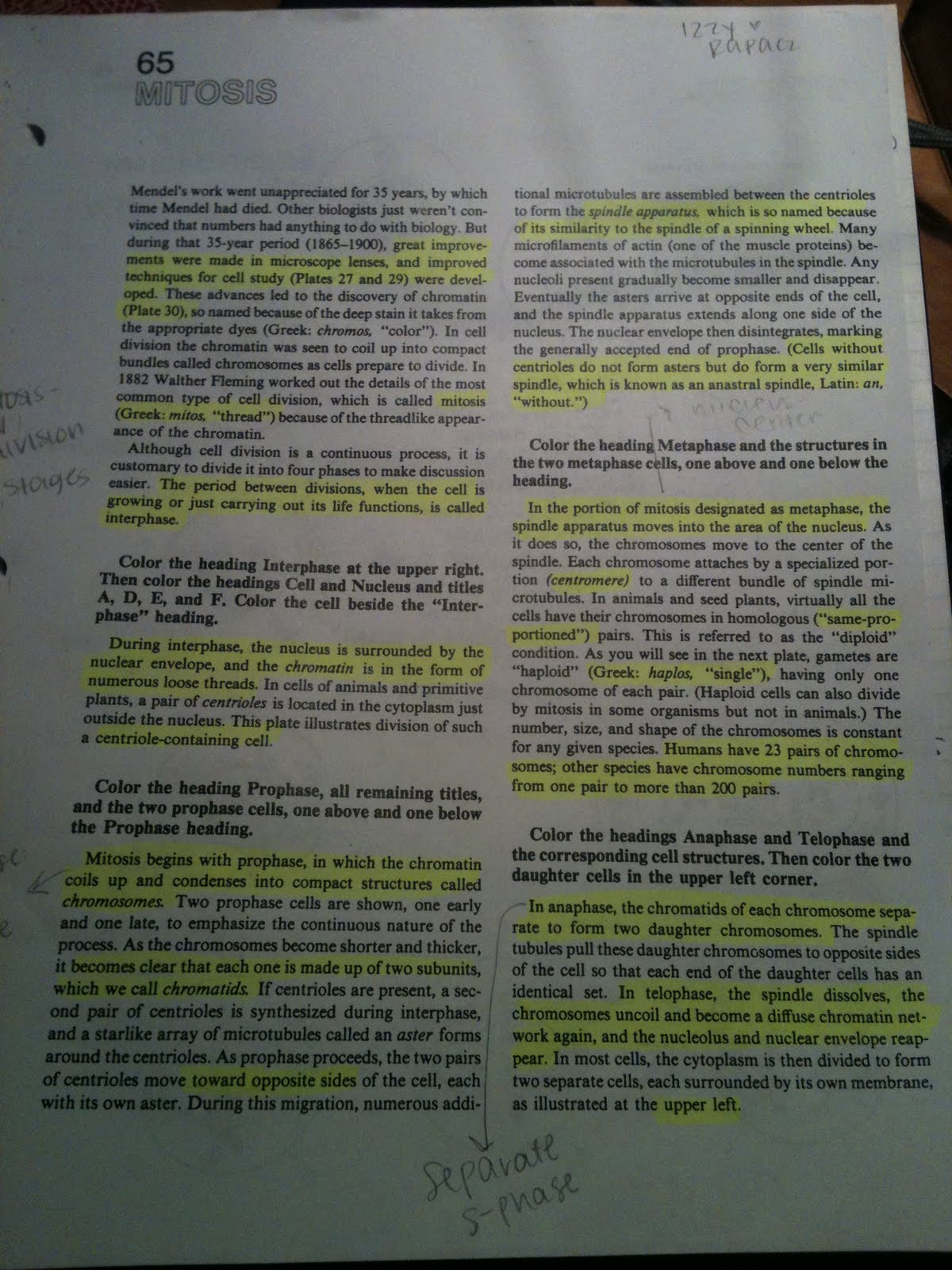
Since today was late arrival with thirty-five minute classes, the class began by walking in, picking up a sheet and a picture, and sitting down. The sheet was our homework, and the picture was to tape into our notes. After organizing ourselves, Mrs. Stein asked for everybody to have their stamp sheet and their Estimation of Cancer Risk Graph located in the UP packet (55). Continuing the morning, Mrs. Stein had notes on the Smart Board for everybody to copy down - there is a picture of them on the left of this scribe. The notes were on the Cell Cycle and basics about it. The cycle has four stages, in which chromosomes take place. The cycle consists of: Interphase (G1), S Phase, G2, and the M Phase. Cells undergo these stages in order to grow, replicate, prepare for mitosis, and finally, divide. Chromosomes are in the cell, and they are the ones that do the jobs. After taking notes for about twenty minutes, we were allowed to work on our Mitosis (65) Worksheet. We had to annotate, and on the back, color the appropriate structures the appropriate color. Since that was fifteen minutes, the bell rang and class
was over. We walked out with our heads filled with science and looking for tomorrow.
HOMEWORK:
1) Annotate the Mitosis worksheet (with notes in the margins!), and color code the back.
2) Start Cancer research.
3) Cancer essay due 10/25.

The Homework. ^ ...... >
Extra Credit!!! Shhhh!!!! It's a secret!

Click on this picture and play the Cell Cycle game! Who is the "supervisor" in the game? Email me the answer! Extra credit will be awarded to the first 5 people from each section who email me the correct answer PRIOR to the start of class. Don't tell anyone. Shhhhh!
Tuesday, October 12, 2010
Enzymes
Here is the picture I showed you in class. It does a nice job illustrating how an enzyme works.


Thursday, October 7, 2010
Plasmolyzed Cell Lab!
Agenda:


1. QUIZ!
2. Bring up 34-35 with stamp sheet
3. Discuss diffusion lab and osmosis in cells
4. Discuss pH enzymes and tomarows lab
5. Plasmolyzed lab (UP 36-37)
Homework:
1. Finish Plasmolysis lab (UP 36-37)
2. Prelab for enzyme lab:
a. Use section 2.4 in textbook for extra help
b. Lab on UP 24-30
When we first got to class we took the quiz. After that we got our stamp for the diffusion lab. Next, as a class we discussed the diffusion lab and answered any questions we had. Mrs. Stein then explained how osmosis in cells works. She explained that a cell might have 99% water and 1% salt in it, while its in a puddle of 6% salt and 94% water. If this happened then the cell would get rid of all the water in it because it moves from high concentration to lower concentration. When this happens, the cell membrane gets smaller and squeezes all the chloroplast into the middle of the cell. Then we witnessed this for ourselves by doing the Normal and Plasmolyzed Cell lab. We ended the class by working on the Analysis questions. If you dont understand this read page 211 in the textbook.
Plasmolyzed Elodea Plant Cell (in salt water):

Elodea Cell (in tap water):

The next scribe will be: Gabby
The Sausage in the Cup
Today we did a lab which was an example of diffusion. diffusion is when one object passes through a cell or a cell membrane into a less densly populated area. The lab that we did involved a plastic tube about 7 inches long, 200ml beaker, glucose solution, an iodine solution, and a soluble starch solution. First we got the plastic tube and tied a string tightly to the bottom of it, then we filled it with the starch solution. After the tube was filled, we added 20 drops of glucose into the starch. Meanwhile the others in the group filled a beaker with water and added a good squirt of iodine into the beaker.
Then we put the plastic tube into the beaker and waited. while we waited we reviewed some of the organells that we have a test on for tomarrow.
When we came back the glucose and the starch had a purpleist tint to it. We later learned that the iodine from the beaked had diffused into the plastic bag. it did this because there was a large population of iodine in the beaker and some of it wanted to get into a less populated area.
That is how the lab went so we cleaned up and and started on answering the questions in the unit packet.
Homework :
-Finish diffusion lab, Pgs.33-35
-STUDY FOR ORGANELL QUIZ!

The next scribe is Natatlie
Then we put the plastic tube into the beaker and waited. while we waited we reviewed some of the organells that we have a test on for tomarrow.
When we came back the glucose and the starch had a purpleist tint to it. We later learned that the iodine from the beaked had diffused into the plastic bag. it did this because there was a large population of iodine in the beaker and some of it wanted to get into a less populated area.
That is how the lab went so we cleaned up and and started on answering the questions in the unit packet.
Homework :
-Finish diffusion lab, Pgs.33-35
-STUDY FOR ORGANELL QUIZ!
The next scribe is Natatlie
Wednesday, October 6, 2010
Lab Questions
Ok. I'm very confused, one of the questions says the starch didn't diffuse and i thought it did. So did it or didn't it???? Help me please!
Tuesday, October 5, 2010
Cells 10/5/10 Scribe Post
Hello everyone!
The next scribe is Joel
We started off class today by turning in our "Check My Understanding" sheet (pages 226-228 + 250-251) which was due today. While we were turning the homework in, we collected two papers from the counter. The large green paper that we got was the "Prelab for Diffusion Lab" and the other small paper was a diagram of cellular respiration. Next, Mrs. Stein showed us the rest of the slide show which gave us information about the mitochondria(converts chemical energy), biochemical energy(cells store and release energy using ATP), cellular respiration(energy fomr glucose is released), lysosomes(breaks materials down into small molecules to be reused), vacuole(stores materials), cytoskeleton(provides support/shape and is involved in movement) and cell walls(provides protection and support). Mrs. Stein said that if you could not finish all the notes, then she would have the slide show posted on the blog. After watching the slide show, Mrs. stein explained the homework to us. Also, Mrs. Stein announced to us that the Cell Quiz will be on THURSDAY instead of Wednesday.
The homework is as follows:
-Read diffusion prelab on pages 33-34 in your Unit Packet.
-Finish color code and annotations
-Read all of 7.3..... If you were in my row, Mirella's row, Dana's row or Amandas row, you were assigned to do ENDOCYTOSIS and FACILITATED DIFFUSION and the rest of the class was assigned to do MOLECULAR TRANSPORT and MITOSIS. (ex. http://gbs-moodle.glenbrook225.org/moodle/file.php/1521/Reading_Strategies/Definition_Mapping.pdf)
-Reminder: there is still time to do the extra credit!
For the remainder of class, we worked on our homework!
Sorry... I wanted to upload pictures but it wouldn't let me :(
The next scribe is Joel
Monday, October 4, 2010
Cells
Hello everyone! Today was pretty much a normal day in Bio...
We started off by turning in and/or getting a stamp the homework from yesterday:
1.) UP pgs. 9-14
2.) The organelle chart (stamp)
Next thing we did was we went over the organelle chart. To make things more clear on the different functions of all the structures in a cell Mrs. Stein showed us a slide show. In the slide show she showed us information about the nucleus, ribosomes, ER, golgi bodies, and chloroplasts. If you missed the slide show or missed something you can find the slide show at the bottom.
For the rest of class we worked on homework or did the microscope test (if it wasn't done already).
The homework is the following:
1.) Finish colorcodes w/ Annotations of readings
2.) Read pgs. 226-228 and 250+251 w/"Check My Understanding"
3.) Quiz Wednesday!
Sunday, October 3, 2010
N/A
Hello.
This is Sam.
This Friday we finished up the lab for the majority of the period. Your groups are the same people that you worked with on the rip-o-meter lab. During the lab Mrs. Stein took students and tested them on how well they did when working with a microscope. Once the period is over we cleaned up after ourselves and left.
For if you didn't finish observations or was absent, here are the things we looked at:

 Frog blood
 Cheek Cells
Homework:
This is Sam.
This Friday we finished up the lab for the majority of the period. Your groups are the same people that you worked with on the rip-o-meter lab. During the lab Mrs. Stein took students and tested them on how well they did when working with a microscope. Once the period is over we cleaned up after ourselves and left.
For if you didn't finish observations or was absent, here are the things we looked at:
 Frog blood
 Cheek Cells
Homework:
- Read 7.2 and Complete the Organelle Chart (Chart available on moodle)
- UP 9-14
- Extra Credit (click on "Extra Credit" tag)
Thursday, September 30, 2010
Scribe Post for 9/30/10
Hey everyone,
Today Mrs. Stein started class as usual by walking around checking in the homework from the previous night. Which was to complete the pre-lab on page 8 in our unit 3 packet and to fill in the double bubble map comparing and contrasting eukaryotes and prokaryotes. Then we reviewed the double bubble maps and added some more categories. After we went over all of this, we started our notes for today's class. The notes we took in class were about cell specialization. This category is about cells developing in different ways to perform different tasks. Cell specialization was about complex organisms that have more specialized cells and the structure of a cell determines the function of it. Then the next category we talked about was major cellular regions. Under this category we talked about plasma membrane (outer boundary of the cell), the nucleus (organelle which contains DNA), and the cytoplasm, a gel-like substance between the cell membrane and the nucleus.
After we took some notes and went over the homework, we started our new lab which is called cell structure and function. Mrs. Stein said that it is very important to read all of the directions and label. In this lab we are observing various cells and structures of plants and animals under the microscope. The first plant we observed was the elodea leaf. To observe any of the cells, you first have to make a wet mount slide out of it and then you can observe it. The leaf was very interesting to observe underneath the microscope. You could actually see some of the organelles and other characteristics that make up a cell.
The second cell we started to view was the lugol's iodine. We weren't able to get that far into it but we were able to view it a little bit. The other cells we will be observing are a human cheek cell, onion cell, and frog blood.
Here are just some views of the cells we will be observing:
Elodea Leaf:


- Complete U.P. 9-14 - Monday
- Read 7.2 and complete the organelle chart - Monday (the chart is also available on the blog)
The next scribe will be Sam
Today Mrs. Stein started class as usual by walking around checking in the homework from the previous night. Which was to complete the pre-lab on page 8 in our unit 3 packet and to fill in the double bubble map comparing and contrasting eukaryotes and prokaryotes. Then we reviewed the double bubble maps and added some more categories. After we went over all of this, we started our notes for today's class. The notes we took in class were about cell specialization. This category is about cells developing in different ways to perform different tasks. Cell specialization was about complex organisms that have more specialized cells and the structure of a cell determines the function of it. Then the next category we talked about was major cellular regions. Under this category we talked about plasma membrane (outer boundary of the cell), the nucleus (organelle which contains DNA), and the cytoplasm, a gel-like substance between the cell membrane and the nucleus.
After we took some notes and went over the homework, we started our new lab which is called cell structure and function. Mrs. Stein said that it is very important to read all of the directions and label. In this lab we are observing various cells and structures of plants and animals under the microscope. The first plant we observed was the elodea leaf. To observe any of the cells, you first have to make a wet mount slide out of it and then you can observe it. The leaf was very interesting to observe underneath the microscope. You could actually see some of the organelles and other characteristics that make up a cell.
The second cell we started to view was the lugol's iodine. We weren't able to get that far into it but we were able to view it a little bit. The other cells we will be observing are a human cheek cell, onion cell, and frog blood.
Here are just some views of the cells we will be observing:
Elodea Leaf:
- Complete U.P. 9-14 - Monday
- Read 7.2 and complete the organelle chart - Monday (the chart is also available on the blog)
The next scribe will be Sam
Hey guys today in class we had an absolute amazing fun time like we always do. The agenda for the day was as following:
1)Grades/Attendence/Star and a Wish
2)New Unit! (cells and cancer)
3)Notes on new unit
4)Rewiew of miccroscope
The homework was as following:
1)Read 7.1 and compplete double bubule map on prokaryotes and eukaryotoes
2)UP page 8
3)answer forum
4)extra credit
After we took attendece we started to discuss our new unit. We took notes on things we already know about cells. Then she gave us a power piont about the levels of organization and the cell theory. I am having some difficulties getting them up on the page so look for them tonight i should have it figured out. Then we went on to practice using the new microscope with our partners. In the next 2 days Mrs.Stien willl be testing us on how well we can use the microscope. Good luck! then from there we packed up the microscope and headed back to our seat where we were dismiss. Thanks for reading!
The next scribe is who ever I piont to in class :p
Subscribe to:
Posts (Atom)